GFP Expressing Human Umbilical Vein Endothelial Cells

Quick Actions
Product Overview
Description
GFP-HUVECs are engineered endothelial cells that preserve essential traits typical of endothelial cells, making them a valuable tool for research. Their unique GFP (green fluorescent protein) labeling facilitates straightforward visualization under fluorescence microscopy, enabling researchers to observe various cellular processes in real time.
This fluorescent signal allows for detailed tracking of cell morphology, migration patterns, proliferation rates, tube formation, and interactions with different cell types, all within two dimensional and three dimensional culture systems.
These versatile cells are particularly well-suited for a wide range of scientific inquiries, including investigations into vascular biology, the mechanisms underlying inflammation, studies on wound healing, modeling angiogenesis, and conducting high content imaging assays. Their ability to mimic natural cellular behaviors and interactions supports advanced research and provides insights into complex biological processes.
IMPORTANT: Quality warranty requires use of NeoBioPharma media. Biohazardous material despite negative pathogen testing. Quality claims must be reported within 1 month.
Key Features
Catalog Number: NBP-001GFP
Product Format: Frozen
Vial Cell Number: > 5x10[5] cells/vial
Passage:1(P1)
Storage: Liquid Nitrogen, Vapor Phase
Intended Use: Research use only (RUO)
Characteristics:
-Human umbilical vein endothelial cell origin
-Stable GFP expression
-Bright green fluorescence under GFP/FITC filter
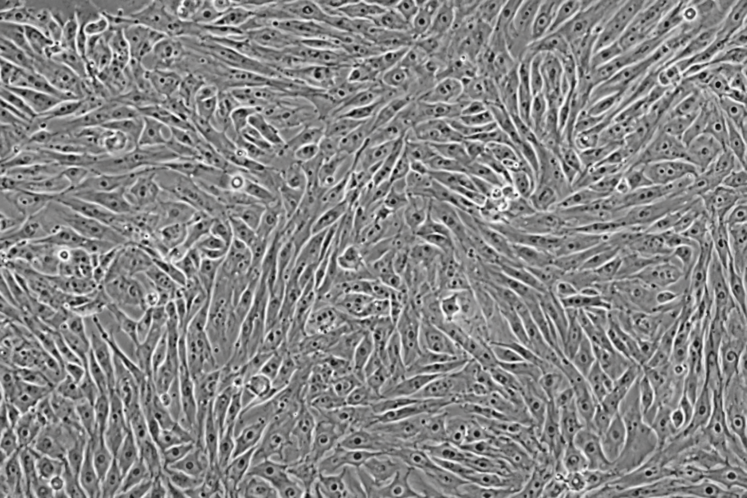
-Endothelial morphology with cobblestone-like appearance
-Suitable for live-cell imaging and fluorescence-based assays
-Express endothelial markers such as CD31, VE-Cadherin, vWF, and VEGFR2
-Capable of migration, proliferation, and tube formation under appropriate conditions
-Research-use-only product
Quick Facts
Product Type
GFP Expressing
Viability
>85% viable (Post-thaw)
Applications
-Angiogenesis and tube formation assays -Endothe…
Availability
In Stock • Global Shipping
Technical Specifications
Primary Applications
Applications & Use Cases
Primary Applications
-Angiogenesis and tube formation assays -Endothelial cell migration studies -Wound healing and scratch assays -Vascular inflammation studies -Drug screening and toxicity testing -Co-culture models with fibroblasts
Suitable for -angiogenesis and tube formation assays -endothelial cell migration studies -wound healing and scratch assays -vascular inflammation studies -drug screening and toxicity testing -co-culture models with fibroblasts studies and research applications
pericytes
Suitable for pericytes studies and research applications
immune cells
Suitable for immune cells studies and research applications
or tumor cells -3D spheroid and organoid vascularization studies -Live-cell fluorescence imaging -High-content screening
Suitable for or tumor cells -3d spheroid and organoid vascularization studies -live-cell fluorescence imaging -high-content screening studies and research applications
Research Areas
In Vitro Studies
Cell culture and screening applications
Molecular Biology
Gene expression & signaling studies
Drug Discovery
Screening & validation studies
Disease Modeling
Pathophysiology research
Support & Resources
Technical Support
Our experts are available for technical questions and protocol assistance.
Contact Support