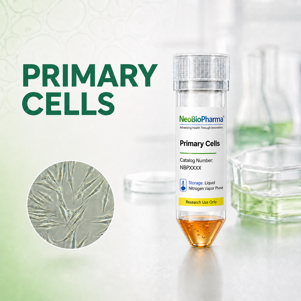
GFP Expressing Human Dermal Fibroblasts-neonatal

Quick Actions
Product Overview
Description
GFP Expressing Human Dermal Fibroblasts – Neonatal are primary-like human skin fibroblasts engineered to stably express Green Fluorescent Protein (GFP). These cells are derived from neonatal dermal tissue and are useful for live-cell imaging, wound healing studies, extracellular matrix research, skin biology, and cell tracking experiments.
Because the cells express GFP, they can be easily visualized under fluorescence microscopy without additional staining.
IMPORTANT: Quality warranty requires use of NeoBioPharma media. Biohazardous material despite negative pathogen testing. Quality claims must be reported within 1 month.
Key Features
Catalog Number: NBP-0008NEOGFP
Product Format: Frozen
Vial Cell Number: > 5x10[5] cells/vial
Passage:1(P1)
Storage: Liquid Nitrogen, Vapor Phase
Intended Use: Research use only (RUO)
Characteristics
Cell morphology: Spindle-shaped, fibroblast-like adherent cells
Fluorescent marker: GFP-positive
Growth type: Adherent monolayer
Species: Human
Tissue source: Neonatal dermis/skin
Quick Facts
Product Type
Human Fibroblasts Cells
Viability
>90% viability post-thaw
Applications
-Wound healing and scratch assays -Skin regenera…
Availability
In Stock • Global Shipping
Technical Specifications
Primary Applications
Applications & Use Cases
Primary Applications
-Wound healing and scratch assays -Skin regeneration studies -Fibroblast migration and proliferation assays -Extracellular matrix production studies -Co-culture with keratinocytes
Suitable for -wound healing and scratch assays -skin regeneration studies -fibroblast migration and proliferation assays -extracellular matrix production studies -co-culture with keratinocytes studies and research applications
melanocytes
Suitable for melanocytes studies and research applications
endothelial cells
Suitable for endothelial cells studies and research applications
or immune cells -3D skin model development -Cell tracking in tissue engineering models -Drug screening and cytotoxicity assays -Fluorescence-based imaging studies
Suitable for or immune cells -3d skin model development -cell tracking in tissue engineering models -drug screening and cytotoxicity assays -fluorescence-based imaging studies studies and research applications
Research Areas
In Vitro Studies
Cell culture and screening applications
Molecular Biology
Gene expression & signaling studies
Drug Discovery
Screening & validation studies
Disease Modeling
Pathophysiology research
Support & Resources
Technical Support
Our experts are available for technical questions and protocol assistance.
Contact Support