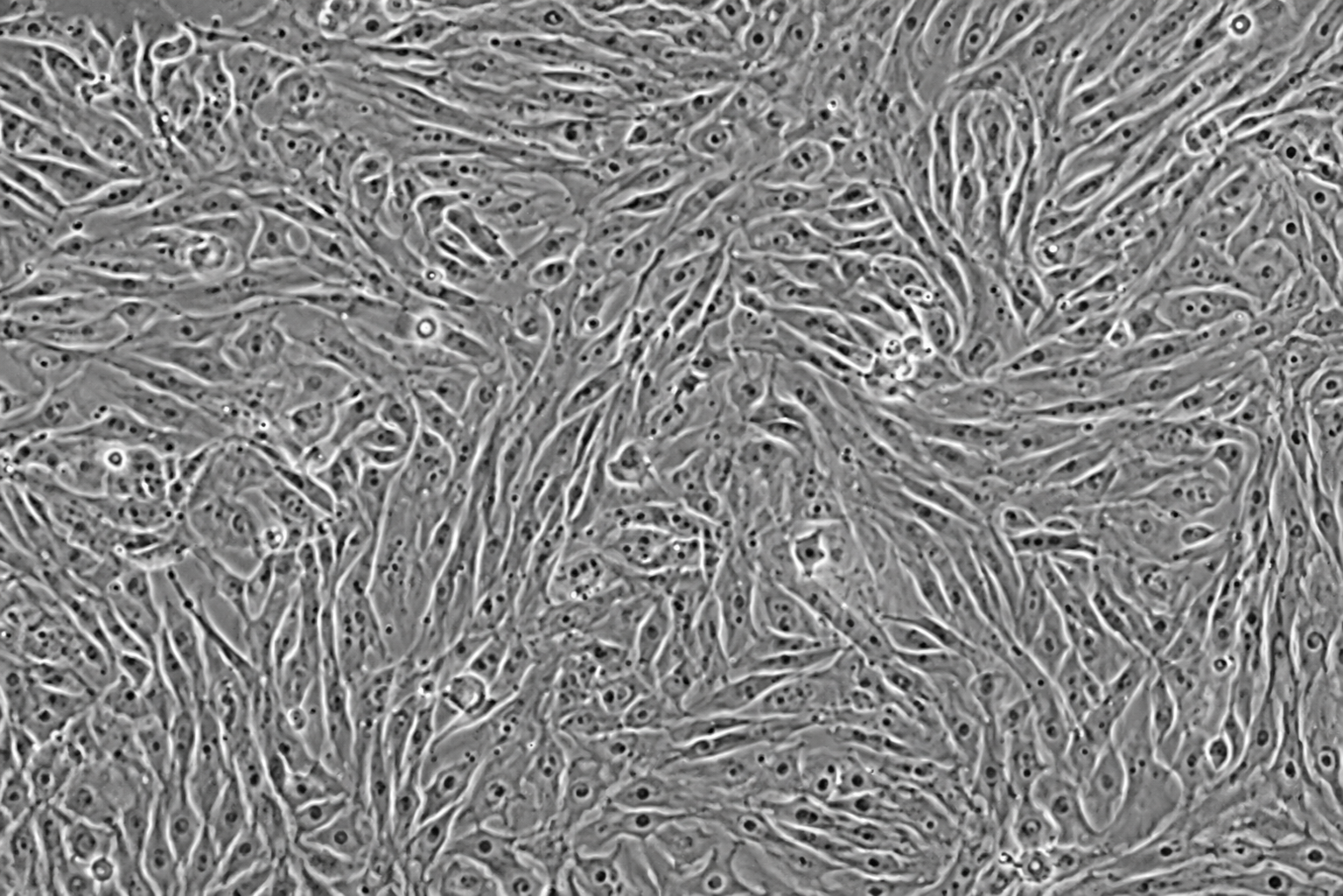
Plasma Membrane GFP Tag Human Umbilical Vein Endothelial Cells

Quick Actions
Documentation 0 files
Product Overview
Description
Plasma Membrane GFP-Tagged Human Umbilical Vein Endothelial Cells are human endothelial cells derived from the umbilical vein and engineered to express green fluorescent protein localized to the plasma membrane. The GFP signal outlines the cell boundary, allowing clear visualization of cell shape, cell–cell junctions, membrane dynamics, and endothelial monolayer organization.
These cells are especially useful for live cell imaging, angiogenesis studies, vascular biology research, permeability assays, and high-content screening, where membrane morphology and endothelial barrier integrity need to be monitored in real time.
Guarantee expansion for a minimum of 2-5 population doublings.
IMPORTANT: Quality warranty requires use of NeoBioPharma media. Biohazardous material despite negative pathogen testing. Quality claims must be reported within 1 month.
Key Features
Catalog Number:NBP-0001GFP-PM
Product Format: Frozen
Vial Cell Number: > 5x10[5] cells/vial
Passage:1(P1)
Storage: Liquid Nitrogen, Vapor Phase
Intended Use: Research use only (RUO)
Characteristics
-Cell type: Human Umbilical Vein Endothelial Cells, HUVECs
-Fluorescent tag: GFP localized to the plasma membrane
-Signal pattern: Bright green fluorescence outlining the cell membrane
-Morphology: Cobblestone-like endothelial morphology under normal culture conditions
-Growth pattern: Adherent monolayer culture
Species: Human
-Tissue source: Umbilical vein
-Functional behavior: Supports endothelial barrier, migration, and angiogenesis-related studies
Quick Facts
Product Type
Human Endothelial cells
Viability
>98% viable (Trypan Blue exclusion)
Applications
1. Live-Cell Imaging Plasma membrane GFP allow…
Availability
In Stock • Global Shipping
Technical Specifications
Primary Applications
Applications & Use Cases
Primary Applications
1. Live-Cell Imaging Plasma membrane GFP allows researchers to visualize endothelial cell borders
Suitable for 1. live-cell imaging plasma membrane gfp allows researchers to visualize endothelial cell borders studies and research applications
cell shape
Suitable for cell shape studies and research applications
spreading
Suitable for spreading studies and research applications
migration
Suitable for migration studies and research applications
and membrane remodeling in real time. 2. Angiogenesis Research Useful for tube formation assays
Suitable for and membrane remodeling in real time. 2. angiogenesis research useful for tube formation assays studies and research applications
endothelial sprouting studies
Suitable for endothelial sprouting studies studies and research applications
and vascular network formation analysis. 3. Endothelial Barrier Studies Ideal for studying cell–cell contact
Suitable for and vascular network formation analysis. 3. endothelial barrier studies ideal for studying cell–cell contact studies and research applications
monolayer integrity
Suitable for monolayer integrity studies and research applications
permeability
Suitable for permeability studies and research applications
and disruption of endothelial barriers. 4. Cell Migration and Wound Healing Assays The membrane GFP signal makes it easier to track individual cell movement
Suitable for and disruption of endothelial barriers. 4. cell migration and wound healing assays the membrane gfp signal makes it easier to track individual cell movement studies and research applications
wound closure
Suitable for wound closure studies and research applications
and collective endothelial migration. 5. Drug Screening and Toxicity Testing Can be used to evaluate how compounds affect endothelial morphology
Suitable for and collective endothelial migration. 5. drug screening and toxicity testing can be used to evaluate how compounds affect endothelial morphology studies and research applications
viability
Suitable for viability studies and research applications
membrane integrity
Suitable for membrane integrity studies and research applications
and vascular function. 6. Inflammation and Vascular Disease Models Useful for studying endothelial responses to inflammatory cytokines
Suitable for and vascular function. 6. inflammation and vascular disease models useful for studying endothelial responses to inflammatory cytokines studies and research applications
oxidative stress
Suitable for oxidative stress studies and research applications
shear stress
Suitable for shear stress studies and research applications
or disease-related stimuli. 7. Co-Culture and 3D Vascular Models Applicable in co-culture systems with smooth muscle cells
Suitable for or disease-related stimuli. 7. co-culture and 3d vascular models applicable in co-culture systems with smooth muscle cells studies and research applications
pericytes
Suitable for pericytes studies and research applications
fibroblasts
Suitable for fibroblasts studies and research applications
immune cells
Suitable for immune cells studies and research applications
or organoid/vascular models.
Suitable for or organoid/vascular models. studies and research applications
Research Areas
In Vitro Studies
Cell culture and screening applications
Molecular Biology
Gene expression & signaling studies
Drug Discovery
Screening & validation studies
Disease Modeling
Pathophysiology research
Support & Resources
Technical Support
Our experts are available for technical questions and protocol assistance.
Contact Support