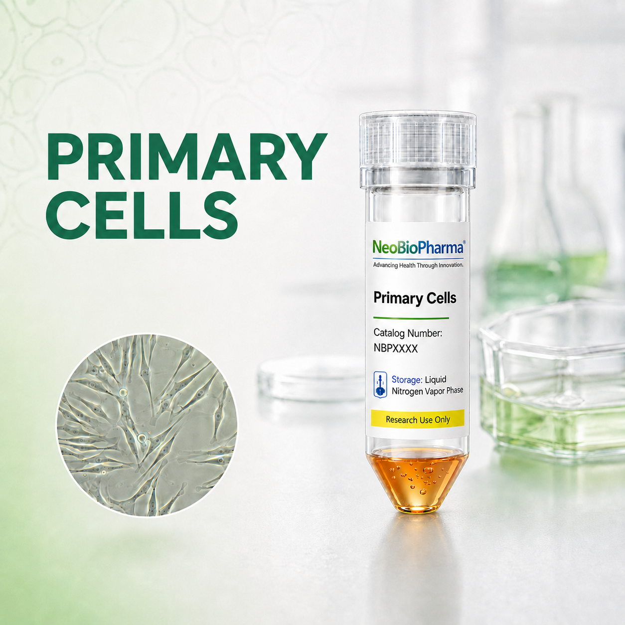
GFP Expressing Human Dermal Lymphatic Microvascular Endothelial Cells

Quick Actions
Product Overview
Description
GFP-Expressing Human Dermal Lymphatic Microvascular Endothelial Cells (GFP-HDLMECs) are specialized primary human lymphatic endothelial cells isolated from the dermal microvasculature of healthy human skin and genetically engineered to constitutively express Green Fluorescent Protein (GFP). These cells retain the biological and molecular characteristics of native lymphatic endothelial cells while providing a stable fluorescent signal that enables real-time visualization and quantitative analysis of cellular behavior.
The lymphatic system plays a critical role in maintaining tissue fluid balance, immune surveillance, lipid transport, and inflammatory responses.
GFP-HDLMECs serve as a powerful in vitro model for studying lymphatic vessel development, lymphangiogenesis, vascular permeability, and interactions between lymphatic endothelial cells and immune, stromal, or tumor cells. Their intrinsic fluorescence eliminates the need for additional labeling procedures, making them particularly valuable for live-cell imaging, time-lapse microscopy, high-content screening, and three-dimensional culture systems.
IMPORTANT: Quality warranty requires use of NeoBioPharma media. Biohazardous material despite negative pathogen testing. Quality claims must be reported within 1 month.
Key Features
Catalog Number: NBP-0003GFP
Product Format: Frozen
Vial Cell Number: > 5x10[5] cells/vial
Passage:1(P1)
Storage: Liquid Nitrogen, Vapor Phase
Intended Use: Research use only (RUO)
Characteristics:
-Cell Type: Human Dermal Lymphatic Microvascular Endothelial Cells (HDLMECs)
-Fluorescent Marker: Stable GFP expression for live-cell imaging
-Morphology: Typical cobblestone-like endothelial morphology when confluent
-Species: Human (Homo sapiens)
-Tissue Origin: Dermal lymphatic microvasculature
-Lymphatic Markers:
LYVE-1 (Lymphatic Vessel Endothelial Hyaluronan Receptor 1)
Podoplanin (PDPN)
PROX1
VEGFR-3 (FLT4)
-Functional Properties:
Tube formation capability
Migration and wound-healing responses
Lymphangiogenic signaling
Barrier function regulation
-Fluorescence: Bright green fluorescence detectable by standard FITC/GFP filter sets
Quick Facts
Product Type
Human Endothelial cells
Viability
>95% viable (Trypan Blue exclusion)
Applications
-Lymphangiogenesis Research Investigation of lym…
Availability
In Stock • Global Shipping
Technical Specifications
Primary Applications
Applications & Use Cases
Primary Applications
-Lymphangiogenesis Research Investigation of lymphatic vessel formation and remodeling Evaluation of VEGF-C/VEGFR-3 signaling pathways Analysis of lymphatic endothelial proliferation and migration -Cancer Research Tumor-associated lymphangiogenesis studies Cancer metastasis through the lymphatic vessels Tumor microenvironment modeling Screening of anti-lymphangiogenic therapeutics -Live-Cell Imaging Real-time monitoring of cell behavior Cell tracking in co-culture systems Time-lapse microscopy experiments Visualization of lymphatic network formation -Drug Discovery Screening compounds affecting lymphatic vessel growth Testing anti-inflammatory and anti-cancer agents Assessment of lymphatic-targeted therapies -Tissue Engineering & Regenerative Medicine Development of engineered lymphatic tissues Vascularized skin substitutes Wound healing and tissue repair studies -Cell Interaction Studies Immune cell trafficking assays Endothelial-stromal interactions Co-culture and organ-on-a-chip models -Advantages of GFP Expression Enables non-invasive visualization without additional staining Supports long-term live-cell tracking Facilitates fluorescence-based quantification assays Improves reproducibility in migration and angiogenesis studies Compatible with high-content imaging platforms -Typical Research Areas Lymphatic biology Lymphedema research Cancer metastasis Inflammation and immune cell trafficking Vascular biology Tissue engineering Drug screening and therapeutic development
Suitable for -lymphangiogenesis research investigation of lymphatic vessel formation and remodeling evaluation of vegf-c/vegfr-3 signaling pathways analysis of lymphatic endothelial proliferation and migration -cancer research tumor-associated lymphangiogenesis studies cancer metastasis through the lymphatic vessels tumor microenvironment modeling screening of anti-lymphangiogenic therapeutics -live-cell imaging real-time monitoring of cell behavior cell tracking in co-culture systems time-lapse microscopy experiments visualization of lymphatic network formation -drug discovery screening compounds affecting lymphatic vessel growth testing anti-inflammatory and anti-cancer agents assessment of lymphatic-targeted therapies -tissue engineering & regenerative medicine development of engineered lymphatic tissues vascularized skin substitutes wound healing and tissue repair studies -cell interaction studies immune cell trafficking assays endothelial-stromal interactions co-culture and organ-on-a-chip models -advantages of gfp expression enables non-invasive visualization without additional staining supports long-term live-cell tracking facilitates fluorescence-based quantification assays improves reproducibility in migration and angiogenesis studies compatible with high-content imaging platforms -typical research areas lymphatic biology lymphedema research cancer metastasis inflammation and immune cell trafficking vascular biology tissue engineering drug screening and therapeutic development studies and research applications
Research Areas
In Vitro Studies
Cell culture and screening applications
Molecular Biology
Gene expression & signaling studies
Drug Discovery
Screening & validation studies
Disease Modeling
Pathophysiology research
Support & Resources
Technical Support
Our experts are available for technical questions and protocol assistance.
Contact Support